Mastering Histology Visual Learning for Medical students
Affiliate link — we may earn a commission. Learn more
Mastering Histology Visual Learning for Medical Students – taught by Mohamed AL ashram – is a comprehensive Udemy course that lets you explore microscopic anatomy without leaving your desk. This free histology course delivers high‑resolution slide images, step‑by‑step explanations, and exam‑ready practice for anyone who wants to learn histology online. Updated July 2026, the program covers every major tissue system, practical staining techniques, and basic histopathology, making it ideal for medical, nursing, and science students seeking certification‑level mastery.
What You'll Learn
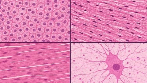
- Build a solid foundation by identifying the four fundamental tissue types—epithelial, connective, muscle, and nervous—on real H&E slides.
- Master cardiovascular, respiratory, and gastrointestinal histology, recognizing organ‑specific microstructures and their functional relevance.
- Learn the complete workflow of tissue preparation, from fixation to embedding, to produce high‑quality microscopic sections.
- Understand staining methods such as Hematoxylin‑Eosin, special dyes, and immunohistochemistry, and how they reveal cellular details.
- Create detailed diagrams of endocrine, reproductive, and urinary tissues, linking morphology to physiological roles.
- Implement basic histopathology principles to spot common pathological alterations in lymphoid and sensory tissues.
- Apply slide‑interpretation strategies that mirror board‑exam questions, boosting confidence for anatomy and pathology assessments.
- Analyze the relationship between tissue architecture and organ function, preparing you for clinical rotations and research projects.
These outcomes ensure you finish the course with practical skills, not just theoretical knowledge.
Course Details
- Instructor: Mohamed AL ashram
- Rating: 4.7 stars (based on student reviews)
- Language: English (en‑GB)
The course is hosted on Udemy, offering on‑demand video lessons that you can pause, rewind, and revisit at any time.
What This Course Covers
Fundamental Tissue Types
- Classification of epithelial tissue: simple, stratified, transitional, and glandular forms.
- Core characteristics of connective tissue, including bone, cartilage, adipose, and blood.
- Overview of muscle tissue types: smooth, skeletal, and cardiac, with functional distinctions.
- Basic organization of nervous tissue, covering neurons, glial cells, and sensory receptors.
Cardiovascular & Respiratory Histology
- Structure of arterial, venous, and capillary walls, highlighting endothelial adaptations.
- Microscopic features of heart muscle layers and conduction system components.
- Airway epithelium variations from nasal passages to alveoli, emphasizing gas‑exchange surfaces.
- Pulmonary vasculature and interstitial tissue relationships in healthy lungs.
Gastrointestinal & Endocrine Systems
- Layered organization of the oral cavity, esophagus, stomach, and intestines, with glandular secretions.
- Histology of liver lobules, pancreatic islets, and adrenal cortex, linking morphology to hormone production.
- Identification of mucosal immune cells within the gut-associated lymphoid tissue.
- Correlation between tissue architecture and digestive enzyme activity.
Urinary, Reproductive & Lymphoid Histology
- Nephron segment identification: glomerulus, proximal tubule, loop of Henle, and collecting duct.
- Male and female reproductive tract histology, focusing on gametogenic and secretory structures.
- Architecture of primary and secondary lymphoid organs, including thymus and spleen.
- Cellular composition of immune zones within lymph nodes and mucosal surfaces.
Histological Techniques & Practical Skills
- Step‑by‑step tissue preparation workflow: fixation, dehydration, embedding, sectioning, and mounting.
- Detailed staining protocols for H&E, PAS, Masson’s trichrome, and immunohistochemical markers.
- Interpretation of staining results, recognizing artefacts versus true pathology.
- Hands‑on virtual lab exercises that simulate slide analysis and report writing.
Each module combines concise video lectures with downloadable slide decks, enabling active learning and quick reference.
Who Should Take This Course
- Medical students preparing for anatomy, pathology, or board examinations.
- Nursing students needing a clear visual understanding of tissue structures for clinical practice.
- Laboratory technicians who perform tissue processing and staining in diagnostic labs.
- Science undergraduates studying physiology, biochemistry, or related fields that require histology knowledge.
- Healthcare professionals transitioning to pathology or research roles that involve microscopic analysis.
Prerequisites
- No prior experience needed — this course is beginner‑friendly and starts with basic concepts.
- Recommended: familiarity with basic cell biology and introductory anatomy terminology.
Why Enroll in This Course
Enrolling now gives you access to a free coupon that provides 100 % off for a limited time, so you can start learning without financial barriers. The course’s visual‑first approach differentiates it from text‑heavy tutorials, allowing you to memorize tissue patterns faster. Because Udemy updates content regularly, you benefit from the latest histology standards and exam‑focused practice.
Course Highlights
- Lifetime access to all video lessons, slide decks, and practice quizzes.
- Self‑paced learning lets you study whenever and wherever you choose, on desktop or mobile.
- Certificate of completion validates your new histology expertise for CVs and résumés.
- High‑resolution slide images provide a realistic laboratory
Affiliate link — we may earn a commission
Affiliate link — we may earn a commission. Learn more